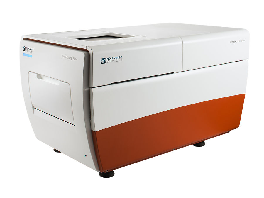
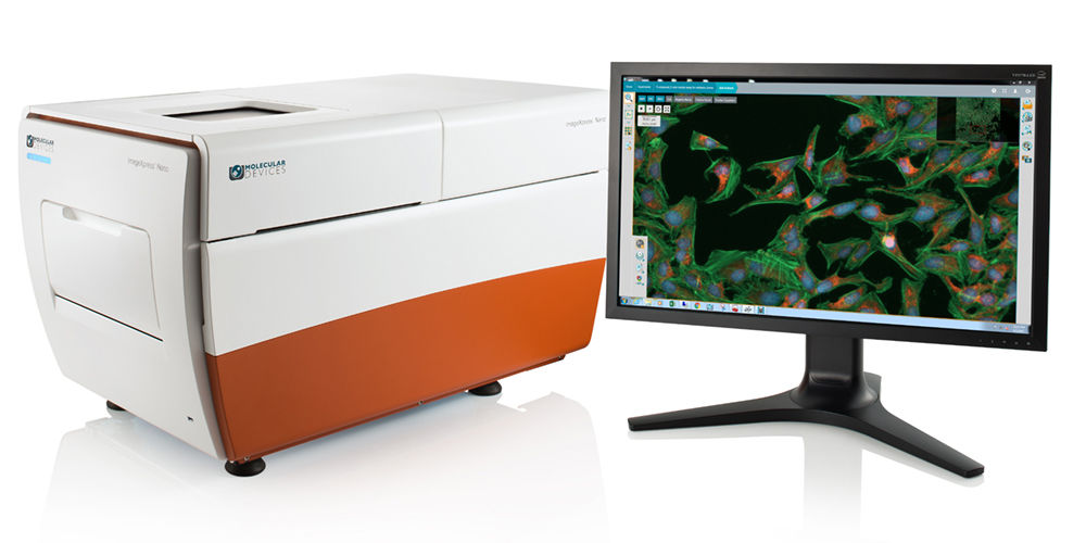
ImageXpress® Nano Automated Imaging System
$undefined
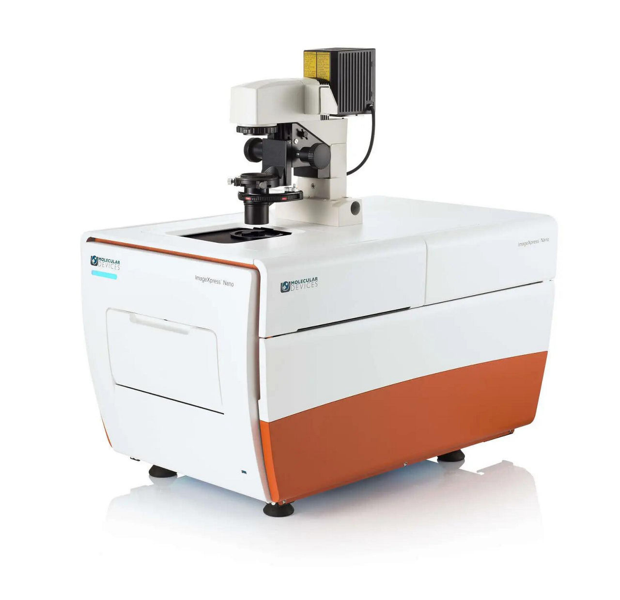
High-content imaging system for your everyday biological needs
Fluorescence imaging platform within reach of every lab
The ImageXpress® Nano Automated Imaging System features a long life, solid state, light engine, and optics to reliably deliver the right assay sensitivity. Capture fine details of a variety of cellular and subcellular assays with this powerful and flexible fluorescent microscopy solution. The system includes MetaXpress High-Content Image Acquisition and Analysis Software with tools for 2D and 3D imaging and time lapse analysis, as well as a range of needs from ease-of-use through to proprietary assay design.
Image label-free
Brightfield imaging allows for rapid acquisition without the use of harmful fluorescent agents.
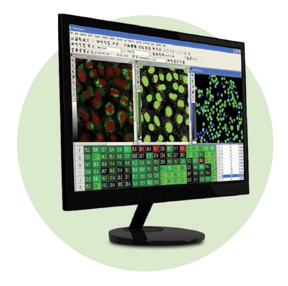
Streamline image analysis
The modular toolbox in the MetaXpress® software allows for the quick setup of hundreds of routine assays. Choose from our optional selection of turnkey application modules for greater convenience.
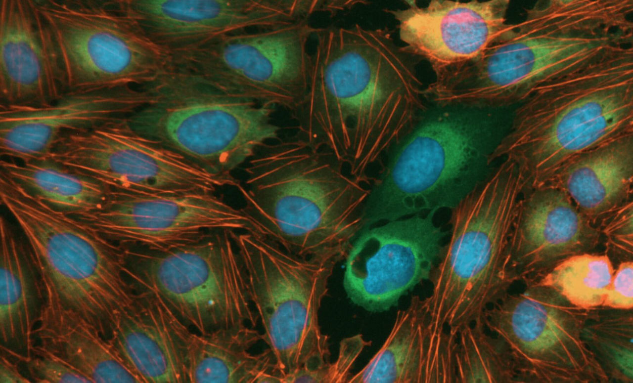
Capture a diverse range of samples
With 2x to 60x magnification, the system offers the flexibility to image whole-well (C. elegans, zebrafish), as well as sub-cellular details (vesicles, organelles).
Large field of view
An entire well of a 384-well plate can be captured in a single image at 4x magnification for faster throughput.
Wide range of filters and objectives
The system can be configured with different filters or objectives (2-60x) to meet research needs.
Automated Stages
Fully automated X, Y, and Z stages with resolution better than 25 nm.
Five fluorescent channels
The system can have up to 5 fluorescent filters installed at one time. The software allows up to 7 channels to be acquired at one time which enables multi-channel fluorescent and transmitted light imaging in one experiment.
High-speed autofocus
Laser autofocus enables quick, consistent focusing across plates, slides and uneven surfaces.
Environmental control option
Multi-day, time lapse and live cell assays can be run using the onboard environmental system with options for temperature, humidity, and CO2 control.
MetaXpress high-content image aquisition and analysis software

- Meet high throughput requirements with a scalable, streamlined workflow
- Adapt your analysis tools to tackle your toughest problems, including 3D analysis
- Schedule automatic data transfer between third-party hardware sources and secure database
- Set up hundreds of routinely used HCS assays using MetaXpress software modules