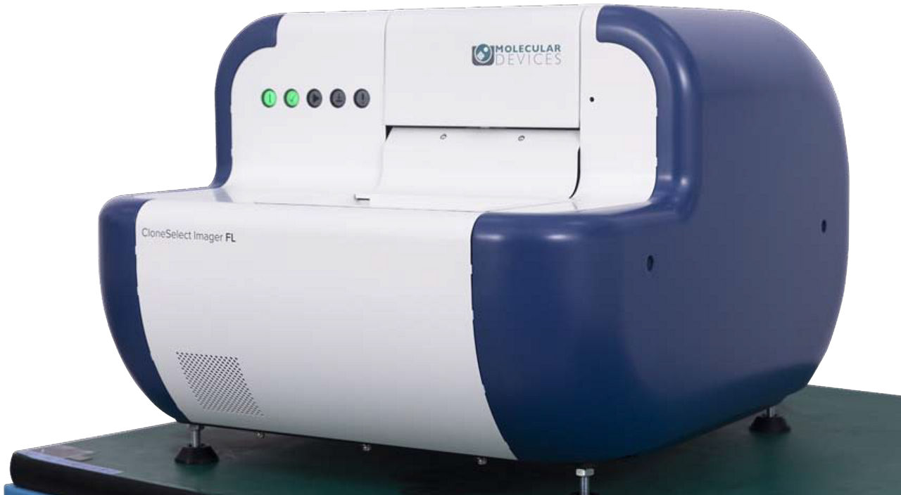
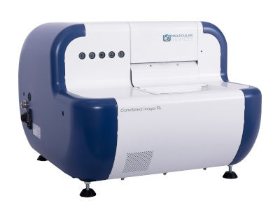
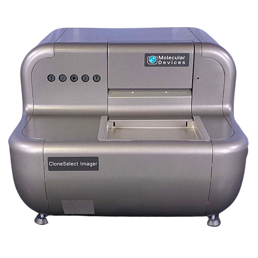
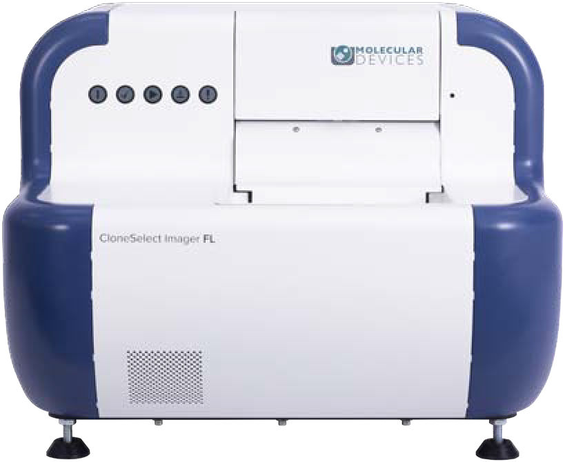
CloneSelect® Imager (CSI and CSI FL)
$undefined

High-speed fluorescence and white light imaging, intelligent data analysis, and monoclonality report generation
Next-generation fluorescent imaging solution for the assurance of monoclonality and automated confluence across diverse cell types
Demonstrating that cell lines are monoclonal – or that a gene was edited as expected – can be a time-consuming and highly-subjective process when relying on conventional technologies. The CloneSelect® Imager and CloneSelect® Imager FL are high-throughput automated solutions for imaging and analyzing mammalian cells. Tracking the formation of a colony from a single cell is effortless as barcoded plates are tracked over time. Automated acquisition and analysis provides accurate, objective, and consistent results.
With high-resolution white light imaging, the CloneSelect Imager provides automated confluence, monoclonality assurance and industry-leading acquisition times with the ability to image a 96-well plate in under two minutes.
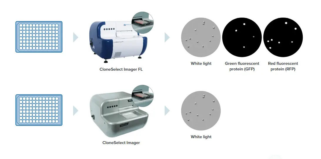
The all-new CloneSelect Imager FL features high-contrast multichannel fluorescent and white light imaging that allows for accurate single-cell detection and proof of monoclonality at day 0. Streamline your workflow with comparative confluence assays to identify and verify gene edits.
Demonstrated IND success
The Monoclonality Report feature streamlines the creation of supporting documentation for regulatory agencies. Reports are automatically generated based on the parameters you select. The Monoclonality Report is an audit-ready document that supports filing for an Investigational New Drug (IND) Application with the FDA. (21 CFR Part 312)
Multichannel imaging and automated confluence
Algorithms are optimized for accurate cell detection and address varying cell types and conditions. Publication-ready high-resolution imaging provides automatic confluence analysis and monoclonality assurance.
Rapid single-cell confirmation
The imager delivers industry-leading acquisition times, imaging a 96-well plate in as little as two minutes and can verify monoclonality from day zero with fluorescence.
White light and fluorescent imaging
Image every well in every plate with rapid acquisition times with label-free white light. Multichannel fluorescence imaging provides additional confidence of monoclonality and comparative confluence assays (red vs green).
Data tools to accelerate research timelines
The software automatically calculates confluence measurements and generates growth curves, heatmaps, and image montages. Measurements for every well are automatically tracked over time. Streamline multiple steps (imaging, sample tracking, data analysis and report generation) in the cell line development workflow for IND filing.
Quickly image a variety of plate formats and cell types
Image a 96-well plate in under two minutes. Compatible with adherent or settled suspension cell types such as CHO, HEK, hybridomas, iPSCs, and many other cell types.
Intelligent analysis with easy-to-use software
The software automatically calculates confluence for each imaging time point. The growth curve, image montage, total growth, and mean rate are generated automatically and are exportable. Guided software user interface allows for simple 1-hour training.
High-resolution images
High-speed fluorescence and high-resolution white light imaging with accurate detection of single cells including debris. Image visualization and data tracking over multiple days.
Custom automation options*
The Automation and Customization Team offers a variety of custom services from robotic plate loading to fully automated workstations with liquid handling and incubation. We have built our custom automated work cells with the CloneSelect Imager for iPSC workflows, full assay systems and drug toxicity and characterization research.
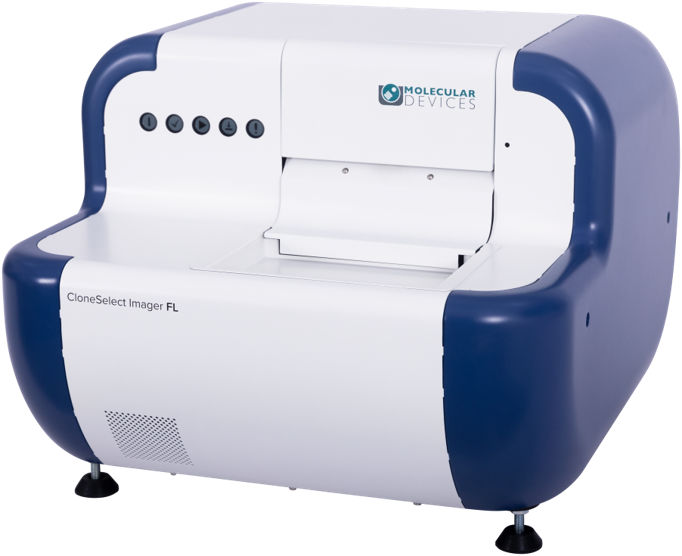
CloneSelect Imager FL
The CloneSelect workflow you know, now with high contrast multichannel fluorescent imaging that allows for verification of gene edits and proof of monoclonality at day zero.
Calculate cell growth curves in every well with multichannel fluorescence and white light imaging across diverse cell types.